Why Study Cell-Cell Interactions?
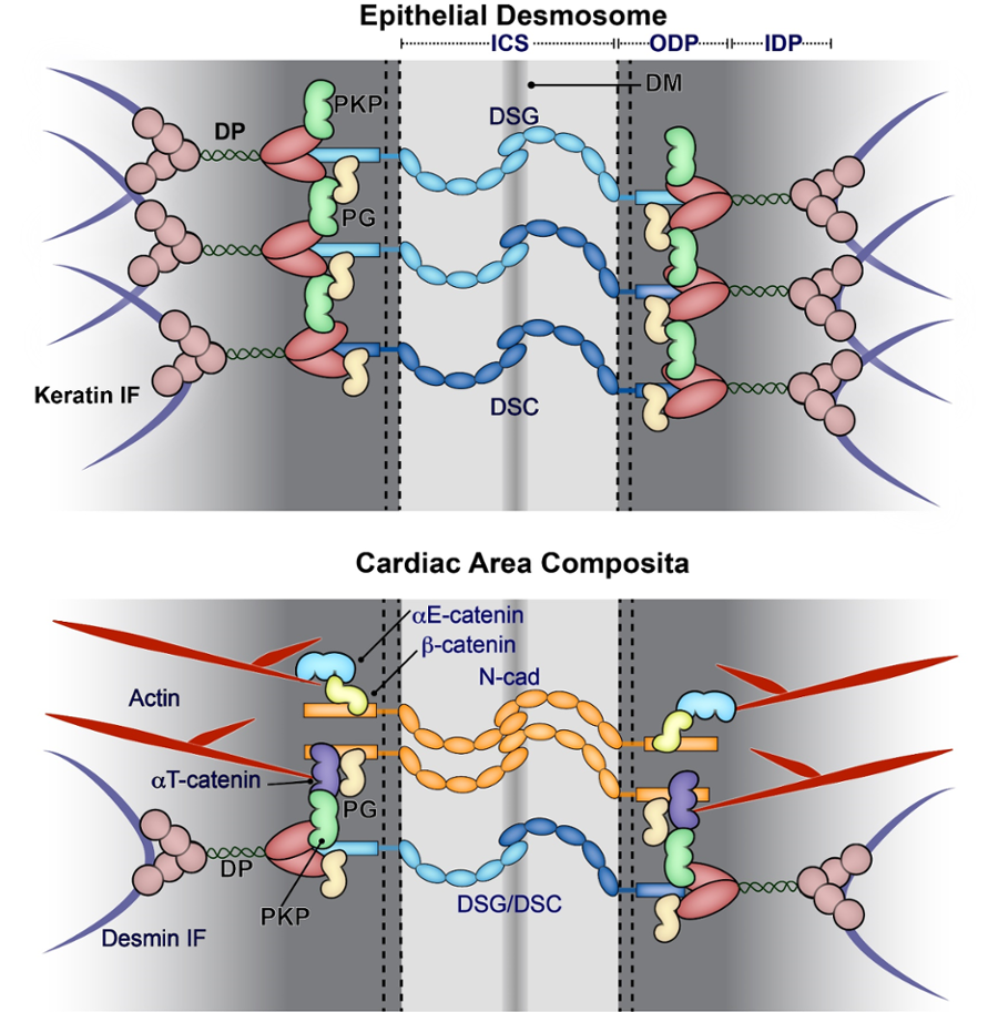
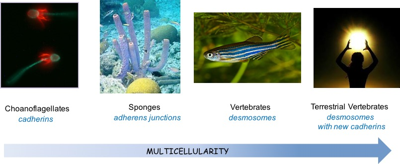
The cell is the fundamental unit of life. An essential property of multicellular organisms is their ability to interact with each other. During evolution organisms co-opted molecules called cadherins, which first appeared in primitive form in single celled choanoflagellates (perhaps functioning to capture prey) to form assemblies called intercellular adherens junctions, which organize the cortical actin cytoskeleton (Fig. 1). Later, in vertebrates, cadherins evolved further and a new type of junction appeared called the desmosome. Desmosomes are molecular assemblies that anchor the intermediate filament cytoskeleton to the cell surface and provide mechanical integrity to complex tissues, particular those under stress such as skin and heart (Fig. 2, 3). Together, these junctions and associated cytoskeletal filaments are essential for normal morphogenetic movements that occur during embryogenesis and organ development, for creating and maintaining cell polarity in differentiated tissues, and for guiding remodeling processes that occur in cancer and other diseases.
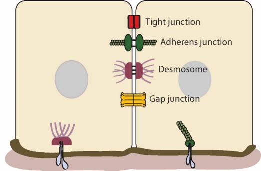
Cell-cell junction functions are not limited to adhesion. Cadherins also engage in bi-directional signaling with growth factor receptors and their downstream signaling effectors: on one hand, activated growth factor receptor tyrosine kinases (RTKs) can negatively regulate cadherin function during the acquisition of cell motility in tumor invasion, while on the other hand, cadherins both positively and negatively regulate the activity of RTKs. They can also serve as transducers of mechanical signaling pathways that control cell behavior. Furthermore, they play a critical role in coordinating the assembly and function of other intercellular junctions such as tight junctions and gap (communicating junctions), which work together to mediate the multiple functions of cell adhesion and communication required in multicellular organisms (Fig. 2). Physical coordination is particularly important in mammalian cardiac myocytes, in which elements of the intercalated disc adherens junctions and desmosomes are intermixed in the area composite (Fig. 3 below).
Finally, our work shows that the evolutionary “newest” desmosomal cadherins can control paracrine signaling in the epidermis through harnessing the production of keratinocyte cytokines and chemokines. Our laboratory studies how cadherins and their associated proteins regulate intercellular adhesion, and also how they integrate cell signaling pathways and cytoarchitecture to regulate cell behavior and tissue morphogenesis.