Defining how cell communication and cooperation drive tissue form and function.
The Green Lab
Just as communication between people is essential for our society to thrive, so too is communication between cells in a multicellular organism essential for its existence. Our group shares a passion for understanding how cells physically stick together to provide mechanical strength to tissues and how adhesion molecules convert mechanical and other environmental cues into signals that drive individual and collective cell behaviors in development, differentiation and disease. We are also passionate about converting our curiosity-driven research into practical knowledge that can help us diagnose and treat adhesion-related diseases, including inherited, autoimmune and bacterial-toxin mediated skin disease, heart disease and cancer. The Green Laboratory is dedicated to an open, collaborative and inclusive research environment promoting high impact research. The lab mentors a diverse team of individuals from different disciplines, backgrounds, perspectives and expertise for a future as independent scientists, educators and professionals in allied fields.
Lab Leadership
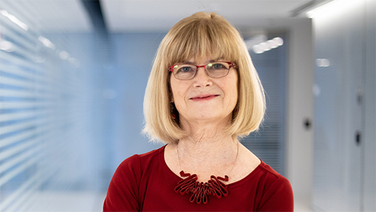
Kathleen J. Green, PhD
Joseph L. Mayberry, Sr., Professor of Pathology
Professor of Dermatology
Associate Director of Basic Sciences, Robert H. Lurie Comprehensive Cancer Center
Lab News
Congratulations to the Green Lab!
Congratulations to Dr. Kathleen Green and the Green Lab, who have received a three-year grant from the LEO Foundation to support their work studying Darier Disease! Thank you to Robert Harmon, Erin McCarthy, Lisa Godsel and Cheryl Olson for their hard work putting together the grant proposal, and their ongoing work studying this rare disease. Read more here.
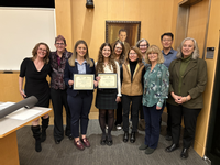
Congratulations Brie!
Our very own Brieanna Jarrell successfully co-organized the 2026 Lurie Cancer Center Nanocourse: “In vitro, in focus: Re-imagining how we study cancer”. Thank you to the excellent speakers and to all who attended!
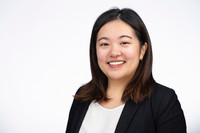
Welcome Farrah!
The Green Lab is thrilled to welcome our new post-doc Dr. Farrah Gao! Farrah recently completed her doctoral training at Case Western Reserve University School of Medicine where she studied how inhibition of the tumor suppressor 15-PGDH protects against injury and disease in the brain and gut, with a particular interest in cellular plasticity and immune crosstalk. She will now be working alongside other Green Lab members to understand how the spatially dependent loss of Dsg1 in melanoma promotes tumor-permissive signals. Welcome to the lab, Farrah!