Research
The Youngblood Lab studies the molecular mechanisms that drive brain tumor growth, aggressiveness and treatment resistance, translating these insights into better therapies for patients. Our projects span the full arc from discovery to translation — defining the genomic and epigenetic drivers of tumor behavior, dissecting how tumors interact with and reshape the immune microenvironment and developing novel therapeutic strategies for tumors that resist surgery. To do this, we integrate a broad range of approaches, including chromatin and epigenomic profiling (e.g., Hi-C, ChIP-seq, ATAC-seq, DNA methylation), spatial and single-cell transcriptomics, immunologic and preclinical modeling and liquid biopsy platforms for tracking tumors over time. Together, these efforts aim to uncover new biology across the spectrum of central nervous system (CNS) tumors and to convert that understanding into meaningful clinical advances.
Current Projects
Advancing Immunotherapeutic Approaches in Meningiomas
Meningiomas are infiltrated by abundant myeloid populations, particularly macrophages, that our lab and others have shown adopt immunosuppressive phenotypes and shape the tumor immune microenvironment. Our program develops preclinical and translational strategies to reprogram these cells from tumor-permissive to tumor-fighting states. A central effort leverages STING pathway activation to reawaken innate immune signaling, while a complementary approach targets the collagen–LAIR1 myeloid checkpoint to relieve inhibitory signaling within the microenvironment. In collaboration with the Dmello Lab, we are also exploiting CHEK2-deficient tumors as a means to remodel the immune landscape and enhance therapeutic response. Together, these projects aim to convert the meningioma myeloid compartment into a driver of anti-tumor immunity.
Representative Papers
- Youngblood MW, Tripathi S, Najem H, et al. STING activation induces cytotoxic and immune responses in meningiomas via inflammatory cell death pathways. Nat Commun. 2026;17(1):2685. Published 2026 Feb 12. doi:1038/s41467-026-69296-1
- Tripathi S, Najem H, Dussold C, et al. Cancer-associated fibroblast-secreted collagen is associated with immune inhibitor receptor LAIR1 in gliomas. J Clin Invest. 2024;134(4):e176613. Published 2024 Feb 15. doi:1172/JCI176613
- Yeung J, Yaghoobi V, Miyagishima D, et al. Targeting the CSF1/CSF1R axis is a potential treatment strategy for malignant meningiomas. Neuro Oncol. 2021;23(11):1922-1935. doi:1093/neuonc/noab075
Key Figure
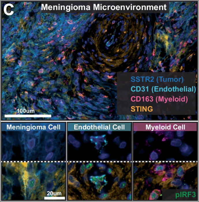
Fluorescence microscopy images of meningioma tissue showing STING protein expressed across neoplastic (SSTR+), endothelial (CD31+) and myeloid (CD163+) populations, with additional images showing activation of the downstream signaling marker pIRF3 in the nucleus of these same cell types.
Source: Youngblood MW, et al. Nat Commun. 2026; PMID 41680157.
Collaborators & Support
Collaborators
- Amy Heimberger, MD, PhD
- Crismita Dmello, PhD
- Rimas Lukas, MD
- Michael Curran, PhD (University of Texas MD Anderson Cancer Center)
- David Raleigh, MD, PhD (University of California San Francisco)
- Stephen Magill, MD, PhD
Support
Decoding the Epigenetic Drivers of CNS Tumor Aggressiveness
Meningiomas and gliomas span a wide clinical spectrum, from indolent lesions cured by surgery to aggressive tumors that recur and resist treatment — yet the molecular basis for this divergence remains incompletely understood. Our lab applies a comprehensive suite of epigenomic technologies, including Hi-C, ChIP-seq, RNA profiling, DNA methylation and spatial epigenetic approaches to define the regulatory programs that drive tumor behavior. By integrating these layers, we aim to identify the epigenetic features that distinguish a benign lesion from one destined to progress despite surgical resection. Ultimately, this work seeks to translate molecular insight into improved prognostication and new therapeutic targets for the most aggressive CNS tumors.
Representative Papers
- Wang Q, Wang J, Mathur R, et al. Spatial 3D genome organization reveals intratumor heterogeneity in primary glioblastoma samples. Sci Adv. 2025;11(11):eadn2830. doi:1126/sciadv.adn2830
- Mathur R, Wang Q, Schupp PG, et al. Glioblastoma evolution and heterogeneity from a 3D whole-tumor perspective. Cell. 2024;187(2):446-463.e16. doi:1016/j.cell.2023.12.013
- Chen WC, Choudhury A, Youngblood MW, et al. Targeted gene expression profiling predicts meningioma outcomes and radiotherapy responses. Nat Med. 2023;29(12):3067-3076. doi:1038/s41591-023-02586-z
- Youngblood MW, Erson-Omay Z, Li C, et al. Super-enhancer hijacking drives ectopic expression of hedgehog pathway ligands in meningiomas. Nat Commun. 2023;14(1):6279. Published 2023 Oct 7. doi:1038/s41467-023-41926-y
- Youngblood MW, Duran D, Montejo JD, et al. Correlations between genomic subgroup and clinical features in a cohort of more than 3000 meningiomas. J Neurosurg. 2019;133(5):1345-1354. Published 2019 Oct 25. doi:3171/2019.8.JNS191266
- Harmanci AS, Youngblood MW, Clark VE, et al. Integrated genomic analyses of de novo pathways underlying atypical meningiomas. Nat Commun. 2017;8:14433. Published 2017 Feb 14. doi:1038/ncomms14433
- Clark VE, Harmanci AS, Bai H, et al. Recurrent somatic mutations in POLR2A define a distinct subset of meningiomas. Nat Genet. 2016;48(10):1253-1259. doi:10.1038/ng.3651
Key Figure
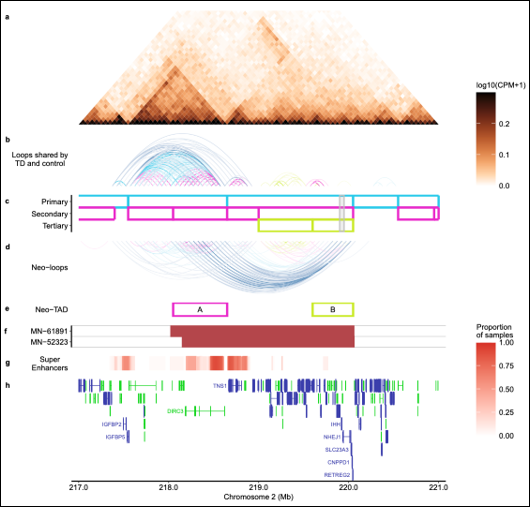
A multi-panel genomic diagram of a region on chromosome 2 showing how a tandem duplication in meningioma tumors rearranges the 3D structure of the DNA, creating new chromatin loops and boundaries ('neo-loops' and 'neo-TADs') that bring enhancer elements near the DIRC3 gene into contact with the IHH gene. Supporting tracks below show the duplication's location, associated super-enhancer activity and nearby genes.
Source: Youngblood MW, et al. Nat Commun. 2023; PMID 3780562.
Collaborators & Support
Collaborators
- Feng Yue, PhD
- David Raleigh, MD, PhD (University of California San Francisco; UCSF)
- Joe Costello, PhD (UCSF)
Support
Tracking Brain Tumor Evolution Through Circulating Extracellular Vesicles
Brain tumors continuously shed extracellular vesicles into the bloodstream, offering a minimally invasive window into tumor biology that conventional imaging and tissue sampling cannot provide. In collaboration with the Nagrath Lab at the University of Michigan, we apply microfluidic immunoaffinity platforms to isolate these tumor-derived vesicles from patient blood using tumor-specific surface markers. By characterizing the molecular cargo of these vesicles, we aim to track how tumors evolve over the course of treatment and to identify early signatures of therapeutic response or resistance. Ultimately, this liquid biopsy approach seeks to illuminate the biology of the tumor microenvironment in real time without the need for repeat surgery.
Representative Papers
- Youngblood MW, Kumari A, Kang YT, et al. Dynamic release of extracellular particles after opening of the blood-brain barrier predicts glioblastoma susceptibility to paclitaxel. Nat Commun. 2025;16(1):11045. Published 2025 Dec 16. doi:1038/s41467-025-65681-4
- Zhang DY, Gould A, Happ HC, et al. Ultrasound-mediated blood-brain barrier opening increases cell-free DNA in a time-dependent manner. Neurooncol Adv. 2021;3(1):vdab165. Published 2021 Nov 12. doi:1093/noajnl/vdab165
Key Figure
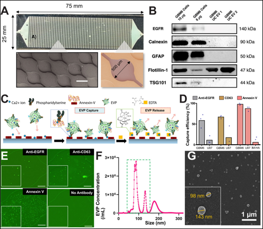
A multi-panel figure showing a microfluidic chip designed to capture tumor-derived extracellular vesicles from blood or cell culture media. Panels show the physical device and its microwell structure, antibody validation by western blot, a diagram of how vesicles are captured and released using surface coatings, bar graphs comparing capture efficiency across different capture methods and cell types, fluorescence images confirming vesicle capture and particle-size measurements confirming the captured vesicles are exosome-sized.
Source: Youngblood MW, et al. Nat Commun. 2025; PMID 4140229762.
Collaborators & Support
Collaborators
- Adam Sonabend, MD
- Sunitha Nagrath, PhD (University of Michigan)
- Abha Kumari (University of Michigan)
Support
Defining Therapeutic Vulnerabilities in Aggressive PitNETs
Pituitary neuroendocrine tumors (PitNETs) cause substantial morbidity through both hormone hypersecretion, which drives systemic endocrine dysfunction and local mass effect, which can produce neurological deficits such as vision loss. Surgery is curative for many patients, but others are left with residual or recurrent disease that remains difficult to control. Our lab applies advanced molecular profiling to define the epigenetic drivers of PitNET aggressiveness and to identify therapeutic vulnerabilities in surgically refractory tumors.
Leveraging Meningioma Biology to Develop Better Therapies
For patients whose meningiomas recur or invade the brain after surgery and radiation, treatment options remain critically limited — there is currently no standard-of-care chemotherapy. Our lab works to close this gap by translating molecular insight into rational therapeutic strategies. In collaboration with the Horbinski and Rogers groups, we are developing local therapy approaches that deliver treatment directly to the surgical resection cavity, concentrating drug at the site of recurrence while sparing systemic toxicity. We have also shown that DNA methylation profiles can nominate effective chemotherapies, identifying docetaxel as an agent that selectively targets high-risk meningiomas and sensitizes them to radiation. Complementing these efforts, our studies of brain invasion biology have revealed the cellular reprogramming and microenvironmental interactions that allow tumors to breach the brain, exposing new vulnerabilities for therapeutic intervention.
Representative Papers
- Aggarwal A, Youngblood MW, Picart T, et al. Meningioma cell reprogramming and microenvironment interactions underlie brain invasion. Neuro Oncol. 2026;28(4):926-938. doi:1093/neuonc/noaf292
- Youngblood MW, Tran AN, Wang W, et al. Docetaxel targets aggressive methylation profiles and serves as a radiosensitizer in high-risk meningiomas. Neuro Oncol. 2023;25(3):508-519. doi:1093/neuonc/noac206
Key Figure
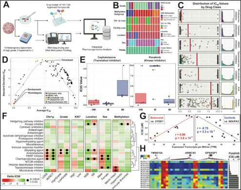
A multi-panel figure showing a drug-screening pipeline that tested 147 FDA-approved compounds against patient-derived meningioma cell cultures to identify effective chemotherapy candidates. Panels show the experimental workflow, patient sample characteristics, the range of drug potency across different drug classes and scatter plots highlighting standout compounds, which include docetaxel, bortezomib and romidepsin, along with charts identifying which patient and tumor factors correlate with drug response.
Source: Youngblood MW, et al. In production.
The image is sourced from this abstract (which matches the study description of 147 FDA-approved compounds), but the image is not included in the paper.
Collaborators & Support
Collaborators
- Craig Horbinski, MD, PhD (Mayo Clinic)
- John Rogers
- David Raleigh (University of California San Francisco; UCSF)
- Stephen Magill