Forebrain and Eye
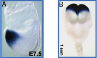
Figure 1. Six3 is one of the earliest markers of the prospective forebrain and eye territory in mammals. A) A E7.5 mouse embryo showing in blue (LacZ staining) the earliest expression of Six3 in the anterior neuroectoderm; B) a few hours later, Six3 expression labels the developing forebrain and eye field.
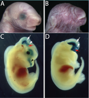
Figure 2. Deletion of Six3 activity arrests forebrain formation in mice. A) newborn WT pup; B) Six3 null littermate lacks anterior structures; C) In an E12.5 Six3 heterozygous embryo, X-gal staining recapitulated the normal pattern of Six3 expression in the eyes, nose (black arrowhead), midbrain, pretectal tegmentum (the white arrow is placed at the boundary between midbrain and pretectum, just caudal to the posterior commissure), ZLI (white arrowhead), and rostral ventral thalamus (red arrow). (D) In the Six3−/− littermates, the residual X-gal staining was detected throughout the midbrain and forebrain tegmentum, the pretectum (weakly stained), and in transverse bands, which may correlate with the ZLI (white arrowhead) and rostral part of the ventral thalamus (red arrow).
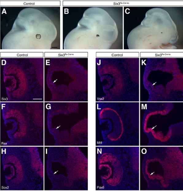
Figure 3. Six3 activity is crucial for the development of the mammalian neuroretina. (A) At E11.5, a normal- looking pigmented eye is seen in control embryos. In Six3 conditional–mutant littermates, the eye appears defective and composed of only the RPE (B and C). At E10.5, NR specification has already taken place in control embryos, as indicated by the expression of the NR markers Six3 (D), Rax (F), Sox2 (H), and Vsx2 (J). In the conditional mutant littermates, this process is defective, as revealed by the lack of expression of any of these NR markers (arrows in E, G, I and K). At this stage, the RPE is also normally detected, as indicated by the expression of Mitf (L) and Pax6 (N). In Six3 conditional–mutant embryos, the expression of these 2 transcription factors highlights the fact that the mutant optic vesicle is exclusively composed of the RPE (arrows in M and O). Scale bars: 500 μm (A–C); 100 μm (D–O).
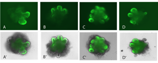
Figure 4. Optic vesicle development in RxGFP cultured stem cells. More recently, we started to take advantage of the 3D organotopic culture system to grow “eyes” in a petri dish. These mini-organs allow us to quickly evaluate candidate genes and substances that could be required during different steps of eye formation. In green (Rx activity), forming optic vesicles can be easily seen.