
Architecture of the filamentous meshwork of the nuclear laminaFigure [a] is a rendered view of a representative 70 nm thick tomogram slice illustrating the putative lamin filaments (yellow), NPCs (red) and occasionally observed cytoplasmic actin filaments (green) on top of the nucleus (n = 55). Scale bar, 200 nm. Figure [b] shows tomogram slices, 2 nm thick, from different regions of the nuclear lamina. Scale bar, 100 nm. Architecture of the filamentous meshwork of the nuclear laminaFigure [a] is a rendered view of a representative 70 nm thick tomogram slice illustrating the putative lamin filaments (yellow), NPCs (red) and occasionally observed cytoplasmic actin filaments (green) on top of the nucleus (n = 55). Scale bar, 200 nm. Figure [b] shows tomogram slices, 2 nm thick, from different regions of the nuclear lamina. Scale bar, 100 nm. 
Vimentin forms a protective cage around the nucleusMaximum projection of Structured Illumination Microscopic images for a vimentin-positive mouse embryonic fibroblast showing that vimentin intermediate filaments (green) assemble into a cage that surrounds the nucleus (magenta). Vimentin forms a protective cage around the nucleusMaximum projection of Structured Illumination Microscopic images for a vimentin-positive mouse embryonic fibroblast showing that vimentin intermediate filaments (green) assemble into a cage that surrounds the nucleus (magenta). 
Vimentin IF template microtubulesQuantitative live-cell imaging of genome edited cells shows that, during directed cell migration, vimentin intermediate filaments assemble in an ultrastructural copy of the polarized microtubule network. Because vimentin turns over slower than microtubules, it stabilizes and templates microtubule organization, thus providing persistence to existing cell polarity. Vimentin IF template microtubulesQuantitative live-cell imaging of genome edited cells shows that, during directed cell migration, vimentin intermediate filaments assemble in an ultrastructural copy of the polarized microtubule network. Because vimentin turns over slower than microtubules, it stabilizes and templates microtubule organization, thus providing persistence to existing cell polarity. 
Vimentin IF are global cytoskeletal elementsImmunofluorescence/phase images of 3T3 cells growing on either square or circular micropatterns having identical areas. Actin microfilaments (MF), vimentin IF (VIF), microtubules (MT) and nuclei (blue) were stained using standard techniques. Averaged cell population heat maps of MF, VIF and MT were generated for square (A) and circle (B) shaped cells. n = number of cells used to generate the heat map. Scale bar is 5 µm. Vimentin IF are global cytoskeletal elementsImmunofluorescence/phase images of 3T3 cells growing on either square or circular micropatterns having identical areas. Actin microfilaments (MF), vimentin IF (VIF), microtubules (MT) and nuclei (blue) were stained using standard techniques. Averaged cell population heat maps of MF, VIF and MT were generated for square (A) and circle (B) shaped cells. n = number of cells used to generate the heat map. Scale bar is 5 µm. 
Organization of lamin isoformsColocalization of lamin isoforms in fibroblasts using indirect immunofluorescence and 3D-SIM. Specific antibodies for pairs of lamin isoforms in all combinations. The results show that lamins A, C, B1, and B2 are each present as distinct meshworks within the nuclear lamina. Organization of lamin isoformsColocalization of lamin isoforms in fibroblasts using indirect immunofluorescence and 3D-SIM. Specific antibodies for pairs of lamin isoforms in all combinations. The results show that lamins A, C, B1, and B2 are each present as distinct meshworks within the nuclear lamina. 
Intermediate filaments in neurodegenerative diseaseGiant Axonal Neuropathy (GAN) is a rare genetic disease in which patients are born normal; but in early childhood they develop severe nerve defects that lead to death at an early age. We demonstrated that the protein mutated in this disease (gigaxonin) is normally involved in the turnover of cytoplasmic IF. While the problem is manifested in nerve cells, patients also have defects in other cells, such as skin fibroblasts, which are much more amenable to cell biological studies. This is illustrated in the image which shows on the left a control fibroblast from an unaffected individual with a normal, well-dispersed IF network and the absence of aggregates. In contrast, the cell on the right from a GAN patient has a large perinuclear aggregate of vimentin. Vimentin is visualized by staining with fluorescent antibodies directed against vimentin. Intermediate filaments in neurodegenerative diseaseGiant Axonal Neuropathy (GAN) is a rare genetic disease in which patients are born normal; but in early childhood they develop severe nerve defects that lead to death at an early age. We demonstrated that the protein mutated in this disease (gigaxonin) is normally involved in the turnover of cytoplasmic IF. While the problem is manifested in nerve cells, patients also have defects in other cells, such as skin fibroblasts, which are much more amenable to cell biological studies. This is illustrated in the image which shows on the left a control fibroblast from an unaffected individual with a normal, well-dispersed IF network and the absence of aggregates. In contrast, the cell on the right from a GAN patient has a large perinuclear aggregate of vimentin. Vimentin is visualized by staining with fluorescent antibodies directed against vimentin.
Laboratory for Cytoskeletal and Nucleoskeletal Dynamics
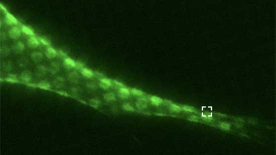
Our lab investigates the structure and function of intermediate filaments (IF), a major cytoskeletal component that plays a critical role in providing structural and mechanical support to the cell. IF are the most diverse of the three major cytoskeletal filaments, being encoded by approximately 70 different genes. This large family of proteins is subdivided into five types, four of which are located in the cytoplasm (cytoskeletal IF) and one type that is restricted to the nucleus (nucleoskeletal IF), and all mammalian cells contain one or more of these IF types. Dysregulation of IFs causes a wide range of human diseases including blistering diseases of the skin, cardiomyopathies, lipodystrophy, muscular dystrophies and neuropathy. The Goldman laboratory provides a supportive, interactive environment in which students and fellows carry out cutting edge research in preparation for careers as independent scientists and educators. Please enjoy perusing our site, and do not hesitate to contact us for further information.

Robert D. Goldman, PhD
Professor
Department of Cell and Developmental Biology
Professor, Department of Medicine-Pulmonary
Northwestern University
Feinberg School of Medicine